Neonatal Lung Abscess-Juniper Publishers
Juniper Publishers-Journal of Pediatrics
Neonatal lung abscess
A baby girl was born at 23+4 weeks by precipitate
delivery following spontaneous onset of labour and PROM of 121 hours.
Mum had received a course of antenatal steroids. Following two
unsuccessful trials of extubation on day 7 and 12, she had a significant
cardio respiratory deterioration on day 14. She was started on second
line antibiotics. Her CRP went up to 20 and her
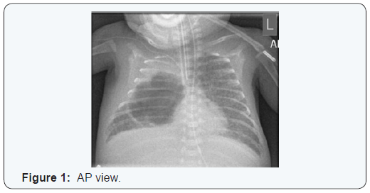
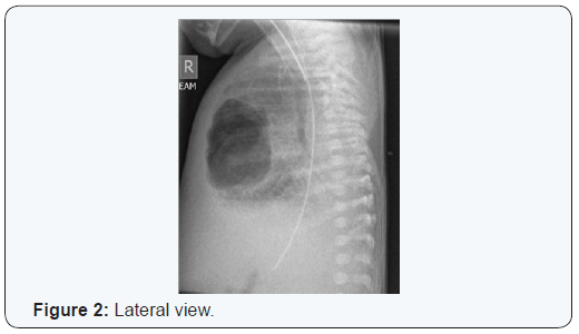
platelet count dropped to 51. Her chest x ray showed a large bullous
emphysematous lesion occupying almost all of her right middle
lobe (Figure 1 & 2). She became increasingly difficult to ventilate
with worsening acidosis, hypotension and hyperglycemia despite
maximizing intensive care support. Following discussion with her
parents, her care was redirected to a palliative course. Post mortem
examination of lungs revealed widespread collections of neutrophils /
abscesses consistent with congenital pneumonia with abscess (seen as
cavitating lesion on imaging). Her blood cultures did not reveal any
growth.
Neonatal lung abscess is very rare [1] and is often
of multibacterial etiology [2,3]. Predisposing factors include
prematurity, assisted ventilation, congenital lung anomaly and
aspiration. Given the range of potential pathogens, direct culture by
percutaneous needle aspiration under either ultrasound [4] or CT
guidance [2] is recommended to direct early appropriate intravenous
medical therapy and hasten recovery, prevent further complications and
obviate the need for surgery [4].
For more articles in Academic Journal of
Pediatrics & Neonatology please click on:
https://juniperpublishers.com/ajpn/index.php
https://juniperpublishers.com/ajpn/index.php


Comments
Post a Comment